The structure and function of the foot can make individuals more susceptible to typical foot issues.
Frequent sources of foot discomfort encompass conditions like plantar fasciitis, bunions, flat feet, heel spurs, mallet toe, Metatarsalgia, claw toe, and Morton’s neuroma. When experiencing foot pain, effective strategies exist to alleviate the discomfort. This article delivers an introduction to foot anatomy and foot-related problems arising from excessive use, injuries, and the natural process of wear and tear.
Anatomy of the foot
Within each of your feet, you’ll find an intricate arrangement of 28 bones, 30 joints, and over 100 muscles, ligaments, and tendons. These elements collaborate harmoniously to fulfill two primary functions:
- Bearing weight
- Propelling forward movement
Furthermore, the foot exhibits flexibility to accommodate uneven terrains while maintaining stability.
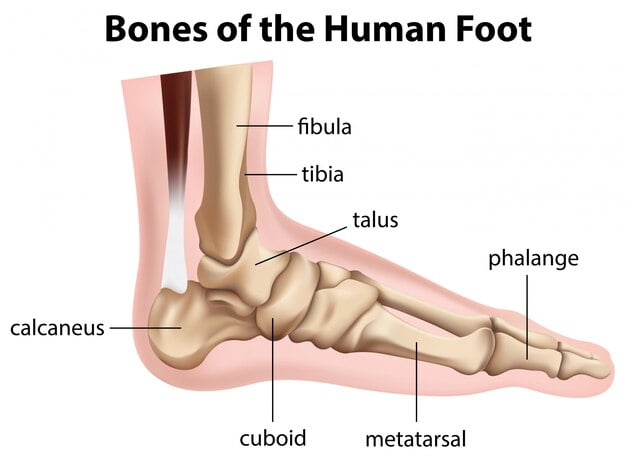
Comprising three distinct sections – the forefoot, midfoot, and hindfoot – each of these segments boasts an array of bones, joints, muscles, tendons, and ligaments.
Foot Bones:
Forefoot Structure
- Phalanges: Comprising the toes, these consist of a total of 14 bones – two for the big toe and three for each of the remaining four toes.
- Metatarsals: These five elongated bones stretch from the base of each toe to the midfoot. The initial metatarsal bone contributes significantly to forward movement, while the second, third, and fourth metatarsals enhance forefoot stability.
- Sesamoid Bones: Positioned beneath the first metatarsal’s plantar surface, two small, oval-shaped sesamoid bones are present. Nestled within a tendon near the bone’s head (closest to the big toe), their purpose is to reinforce the tendon and alleviate stress.
Bones of the Midfoot
Within the midfoot region lie five uniquely contoured bones known as tarsals. Collectively, these tarsal bones shape the foot’s arch, a vital component for bearing weight and ensuring foot stability.[1]
This group of bones comprises:
- Navicular
- Cuboid
- Medial cuneiform
- Intermediate cuneiform
Bones in the Midfoot Region
Situated in the midfoot area are five uniquely shaped bones referred to as tarsals. Collectively, these tarsal bones come together to shape the foot’s arch, a pivotal element for supporting weight and ensuring foot stability.1
This set of bones encompasses:
- Navicular
- Cuboid
- Medial cuneiform
- Intermediate cuneiform
- Lateral cuneiform
Hindfoot Components
Calcaneus: Recognized as the sizable heel bone, the calcaneus assumes a central role by transferring a significant portion of the body’s weight from the legs to the ground.
Talus: Positioned between the calcaneus and the lower leg’s two bones (the tibia and fibula), the talus facilitates the distribution of weight and pressure through the ankle joint.
Muscular Control
The foot’s motion is governed by muscles originating in the lower leg and connected to the foot’s bones via tendons.
The key muscles orchestrating foot movement include:
- Tibialis posterior: This muscle upholds the foot’s arch.
- Tibialis anterior: Enabling upward foot movement.
- Peroneus longus and brevis: Dictating movement along the exterior of the ankle.
- Extensors: Responsible for lifting the toes to facilitate stepping.
- Flexors: Tasked with steadying and curling the toes underneath.
Joints
Joints serve as points of connection between two bones. Within the feet, the big toe comprises two joints: the metatarsophalangeal joint situated at the toe’s base and the interphalangeal joint located just above it.
Conversely, the remaining four toes possess three joints each: the metatarsophalangeal joint at the toe’s base, the proximal interphalangeal joint in the toe’s middle, and the distal phalangeal joint at the toe’s tip.
Tendon
Tendons, comprising tough connective tissues, link muscles to bones. Three primary tendons contribute significantly to foot mobility, facilitating both flexion (forward bending of the foot) and dorsiflexion (backward bending of the foot):
- Achilles Tendon: The most prominent foot tendon, it spans from the calf muscle to the heel. As the body’s largest and strongest tendon, it enables activities such as running, jumping, stair climbing, and rising onto tiptoes.[2]
- Tibialis Posterior: Attaching the calf muscle to the inner foot bones, this tendon supports the foot’s arch.
- Tibialis Anterior: Extending from the outer lower leg bone to the tarsals and first metatarsal, this tendon plays a vital role in dorsiflexion.
Connective Tissues: Ligaments
Ligaments, characterized by resilient fibrous structures, establish connections between bones. The key ligaments within the foot are as follows:
- Plantar Fascia: This extensive ligament spans from the heel to the toes, crafting the arch. It bolsters walking, strength and contributes to equilibrium.[.]
- Plantar Calcaneonavicular: Linking the calcaneus to the talus, this ligament sustains the talus’s head.
- Calcaneocuboid: Bridging the gap between the calcaneus and tarsal bones, this ligament augments the plantar fascia’s role in upholding the foot’s arch.
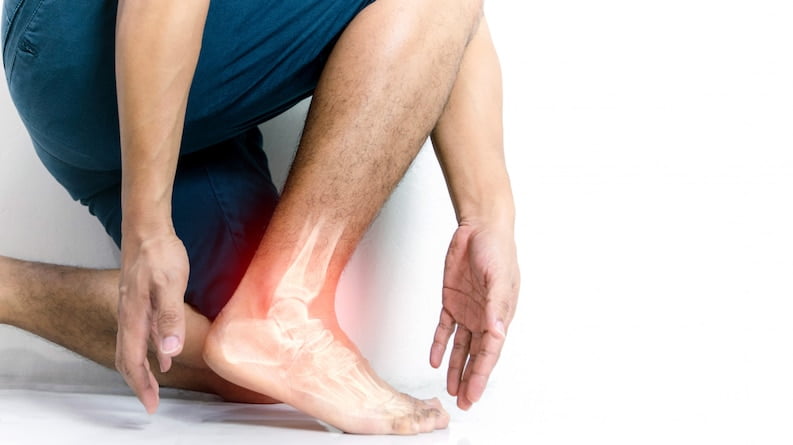
Common Foot Issues
By the age of 50, an individual typically covers around 75,000 miles on foot. Considering the intricate components within the foot, its susceptibility to injury or strain is understandable.[3]
Nine distinct foot-related conditions can induce discomfort, limit foot mobility, or contribute to instability.
Plantar Fasciitis
Microtears in the dense fibrous tissue beneath the foot lead to plantar fasciitis, typically resulting from excessive stretching. Signs encompass heel and arch pain, frequently intensified during mornings. This condition is prevalent among long-distance walkers or runners.[4]
Bunions
Bunions emerge as bony projections either along the inner edge of the foot or the outer side of the pinkie toe. Over time, these develop when foot bones lose alignment, often due to wearing ill-fitting or overly constrictive shoes.
In some cases, the big toe may curve excessively inward, crossing beneath or over the neighboring toe, leading to an additional misalignment termed a hammertoe. Typically, a painful callous tends to develop atop the second toe.
Flat Feet
Flat feet, medically known as PEs planus, occurs when the natural arch of the foot diminishes, sometimes to the extent that the entire sole touches the ground.[5]
This condition can induce discomfort in the midfoot region and lead to ankle and arch swelling. Additionally, the imbalance may contribute to pain in the hip, knee, or lower back.
While pes planus can be congenital, often present from birth, it frequently arises due to aging or injury. Approximately 20% to 30% of individuals exhibit varying degrees of flat-footedness.
Claw Toe
A claw toe is a deformity characterized by a toe bending downward at its middle joints, occasionally even curling beneath the foot. This positioning can lead to the development of calluses or corns on the upper surface of the impacted toe. In certain instances, a corn may exert pressure on nerves within the foot, resulting in discomfort. Numerous approaches for corn removal are available, including at-home solutions.[6]
Mallet Toe
In the mallet toe, the middle joint of a toe undergoes a permanent downward bending, creating a distinctive angle.
This condition arises from an imbalance within the muscles, tendons, or ligaments responsible for maintaining bone alignment. Similar to bunions and hammertoe, mallet toe frequently emerges due to the use of ill-fitting footwear, although it can also stem from injuries or specific medical conditions.[7]
Health Issues That May Lead to Foot Issues
A range of medical conditions can affect the feet, encompassing:
- Sprains and strains
- Ruptured tendons or ligaments
- Osteoarthritis (degenerative joint condition)
- Onychomycosis (nail fungal infection)
- Diabetes-related concerns
- Bone fractures
- Tendinitis (inflammation of tendons)
- Rheumatoid arthritis (autoimmune joint inflammation)
- Gout
- Athlete’s foot
Diagnostic Process
For issues related to the foot’s anatomical components, your healthcare provider or orthopedic specialist will carefully examine your foot, assessing signs of swelling, deformities, skin anomalies, or misalignments.
They will evaluate your symptoms and medical history, observing your walking pattern to detect any irregularities in your gait, a process known as gait analysis.
Diagnosis often involves essential imaging tests, which may encompass:
- X-ray: This standard imaging procedure employs minimal radiation exposure and is effective for identifying conditions such as bone fractures, dislocations, or signs of arthritis damage.
- Computed Tomography (CT): Utilizing multiple X-rays, this imaging method produces a three-dimensional portrayal of the foot’s structure.
- Magnetic Resonance Imaging (MRI): Employing a strong magnet and radio waves, MRI generates highly detailed images without radiation. It excels at visualizing soft tissues.
Treatment Approaches
The course of treatment for a foot ailment hinges on its underlying cause.
Foot discomfort, irrespective of its origin, can often be alleviated with non-prescription pain relievers like Tylenol (acetaminophen), Advil (ibuprofen), or Aleve (naproxen).
In more pronounced instances, the administration of steroid injections might be necessary to diminish joint inflammation, or prescription pain medications like Celebrex (celecoxib) could be prescribed for chronic arthritis pain relief.
Anatomical deformities prompting foot problems, including the presence of a black line on nails, may find relief through the use of foot orthotics (insoles worn within shoes) to mitigate the issues and alleviate pain. While standard versions are accessible at pharmacies, healthcare professionals often prescribe tailor-made orthotics or custom-fitted shoes.
Enhancing foot and ankle strength and flexibility can be achieved through physical therapy. On occasion, conditions such as displaced fractures, bunions, or hammertoes may necessitate surgical intervention if they lead to substantial discomfort or hinder mobility.
Conclusion
The foot is a complex part of our body, made up of bones, muscles, and other parts. It can get hurt from accidents, too much use, or sickness. Some common foot problems include bunions, claw toes, flat feet, hammertoes, heel spurs, mallet toes, metatarsalgia, Morton’s neuroma, and plantar fasciitis.
To find out what’s wrong, a doctor might look at your foot, ask about how you feel, and how you move. They might also take pictures of your foot, like X-rays, CT scans, or MRIs.
How to help depends on the problem. You might need special shoes, exercises, or medicine. Sometimes, if the problem is really bad, you might need an operation.